Kamis, 03 November 2011

Temporomandibular Disorders Lecture note on Oral Medicine


TMJ Disorders
Temporomandibular joint and muscle disorders, commonly called “TMJ” or TMD are a group of conditions that cause pain and dysfunction in the jaw joint and the muscles that control jaw movement.

For most people, pain in the area of the jaw joint or muscles does not signal a serious problem. Generally, discomfort from these conditions is occasional and temporary, often occurring in cycles. The pain eventually goes away with little or no treatment. Some people, however, develop significant, long­term symptoms.

What are the signs and symptoms?
  • Radiating pain in the face, jaw or neck
  • Jaw muscle stiffness 
  • Limited movement or locking of the jaw
  • Painful clicking, popping or grating in the jaw joint when opening or closing the mouth 
  • A change in the way the upper and lower teeth fit together.
What is the Temporomandibular Joint? 
 
The temporomandibular joint connects the lower jaw (the mandible) with its condyle, to the bone at the side of the head—the temporal bone. If you place your fingers just in front of your ears and open your mouth, you can feel the joints.
Because these joints are flexible, the jaw can move smoothly up and down and side to side, enabling us to talk, chew and yawn. Muscles attached to and surrounding the jaw joint control its position and movement.

To keep this motion smooth, a soft disc lies between the mandibular condyle and the temporal bone.
This disc absorbs shocks to the jaw joint from chewing and other movements.

What are TMJ Disorders?
TMJ disorders fall into three main categories:  
  • Myofascial pain, the most common temporo- mandibular disorder, involves dis­comfort or pain in the muscles that control jaw function.
  •  Internal derangement of the joint involves a displaced disc, dislocated jaw, or injury to the condyle.
  •  Arthritis refers to a group of degenerative or inflammatory joint disorders that can affect the temporomandibular joint
A person may have one or more of these conditions at the same time. Some people have other health problems that co­exist with TMJ disorders, such as chronic fatigue syndrome, sleep disturbances or fibromyal­gia, a painful condition that affects muscles and other soft tissues throughout the body.

How jaw joint and muscle disorders progress is not clear. Symptoms worsen and ease over time, but what causes these changes is not known.
Most people have relatively mild forms of the disorder. Their symptoms improve significantly, or disappear spontaneously, within weeks or months.
For others, the condition causes long­term, persistent and debilitating pain. The condition is more common in women than in men.

Prosthodontic treatment, in order to stabilize the occlusion as a result of a TMD articular diagnosis such as localized osteoarthritis or degenerative joint disease (DJD), may be required once the condition has been successfully stabilized. 
The degenerative process creates a smaller condyle which often alters the jaw posture causing an uneven bite.  Thus, it may become necessary to re-establish a more stable occlusion as a result of changes within the joint.

Trauma to the jaw or temporomandibular joint plays a role in some TMJ disorders. But for most jaw joint and muscle problems, scientists and clinicians don’t know the causes.
There is no scientific evidence that clicking sounds in the jaw joint lead to serious prob­lems. Jaw noises alone, without pain or limited jaw movement, do not indicate a TMJ disorder and do not always indicate that treatment is needed.

What causes TMJ Disorders?
The roles of stress and tooth grinding as major causes of TMJ disorders are also unclear. Many people with these disorders do not grind their teeth, and many long­time tooth grinders do not have painful joint symptoms.
Scientists and clinicians note that people with sore, tender chewing muscles are less likely than others to grind their teeth because it causes pain.
Stress may play a role in many persons with jaw joint and muscle disorders that is more likely the result of dealing with chronic jaw pain or dysfunction than the cause of the condition.

How are TMJ Disorders Treated?
Because more studies are needed on the safety and effectiveness of most treatments for jaw joint and muscle disorders, experts recommend using the most conser­vative and reversible treatments when possible.
Reversible treatments do not cause permanent changes in the structure or posi­tion of the jaw or teeth. Even when TMJ dis­orders have become persistent, most patients still do not need aggressive types of treatment.

Treatment by a Prosthodontist may be needed for other reasons such as to restore severely worn, damaged, or diseased teeth or to replace teeth for the purpose of improving chewing, providing enhanced support for your lips or cheeks, or improving the appearance of your smile.
Extensive prosthodontic treatment should only be provided after the TMJ disorder has been adequately diagnosed and its pain successfully managed.

Conservative Treatments
Most jaw joint and muscle problems are temporary and do not get worse. Treatment is based on a proper diagnosis which should be conservative and reversible.
Self-Care Practices
Pain Medications
Stabilization Splints
Prosthodontic Treatment

Self-Care Practices 

Your Prosthodontist may recommend steps that you can take that may be helpful in easing symptoms, such as:
  • eating soft foods,
  • applying ice packs to recommended areas,
  • avoiding extreme jaw movements (such as wide     yawning, loud singing, and gum chewing),
  • learning techniques for reducing stress,
  • practicing gentle jaw stretching and relax­ing exercises that may help increase jaw movement.
Pain Medications

For many people with TMJ disorders, short­-term use of over-­the-­counter pain medicines or nonsteroidal anti-­inflammatory drugs (NSAIDS), such as ibuprofen, may provide temporary relief from jaw discomfort.
When necessary, your dentist or doctor can prescribe stronger pain or anti­inflammatory medications, muscle relaxants, or anti­depressants to help ease symptoms.

Stabilization Splints

Your Prosthodontist may recommend an oral appliance, also called a stabilization splint or bite guard, which is a plastic guard that fits over the upper or lower teeth. Stabilization splints are the most widely used treatments for TMJ disorders.
If a stabilization splint is recommended, it should be used only for a short time and should not cause permanent changes in the way your teeth bite together when the splint is removed from your mouth.

Prosthodontic Treatment


Occlusal splints may also be used to reestablish the bite prior to prosthodontic treatment.
It is used when the bite is not contacting evenly due to missing or worn teeth and may relax the muscles.




Rabu, 02 November 2011

Management of facial trauma-Mid-face Injury-Oral and Maxillofacial Surgery


Mid-face
Definition: 
The area between a superior plane drawn through the zygomatico-frontal sutures tangential to the base of the skull and inferior plane at the level of the maxillary dental occlussal surface.


Structures connection
(structures in relation)
  • Orbit
  • Maxillary sinus
  • Nasal bone
  • Naso-orbital ethmoid (NOE) complex
  • Zygomatic complex
  • Frontal bone and sinus
  • Vertical and horizontal pillars

Area of strength
  • Vertical and horizontal pillars
  • Muscular attachment
Area of weakness
  • Sutures
  • Lining tissues and air-filled cavities
Pattern of fractures of mid-face skeleton
  • Alveolar fracture and dental fracture
  • Le Fort ‘s fracture (french surgeon Rane Le Fort 1901)
  • Naso-orbital ethmoid fracture
  • Zygomatic complex and arch fracture
  • Frontal sinus and bone fracture
Alveolar bone fracture
Involve block of alveolar bone with or without
  • Intrusion of teeth
  • Extrusion of teeth
  • Luxation of teeth
  • Fracture of teeth


Le Fort’s fractures
  • Le Fort I (low level or Guerian fracture)
  • Unilateral/ bilateral
  • Horizontal fracture through the maxilla above the level of the nasasl floor and alveolar process
  • Piriform rims
  • Anterior maxilla
  • Zygomatic buttresses
  • Ptrygoid laminae
Signs and symptoms
  1. Slight swelling of upper lip
  2. Ecchymosis in upper lip sulcus
  3. Hematoma intra-orally over zygoma and in palate
  4. Disturbed occlusion
  5. Mobility of teeth of the involved segment of maxilla
  6. Combination of soft tissue laceration
  7. Exposure of nares and the maxillary antra in case of gross injury
  8. Impacted type of fracture is oftenly not mobile and teeth cusps may be damaged
  9. Cracked-pot percussion of upper teeth
Le Fort II (pyramidal or subzygomatic)

Separation of NF suture, medial orbital walls (lacrimal bone), inferior orbital floor and rim (adjacent to infrorbital canal and foramen), anterior maxilla below zygomatic buttress and ptrygoid laminae about halfway up.
Separation of the block from the base of skull is completed via the nasal septum and may involve the floor of the anterior cranial fossa


LeFort III (cranifacial dysjunction, high transverse, suprazygomatic)
Separation of NF suture, medial orbital walls (involve the depth of the ethmoid bone and cribriform plate, pass below optic foramen and cross the inferior orbital fissur), inferior orbital floor, lateral orbital wall, ZF suture, zygomatic arch, suprazygomatic to the root of ptrygoid plate.
Signs and symptoms
although it is possible to distinguish between le fort II and III, the signs and symptoms are almost similar
  • Gross edema of soft tissue
  • Bilateral circumorbital ecchymosis
  • Bilateral subconjunctival hemorrahge
  • Obvious deformity of the nose
  • Nasal bleeding and obstruction
  • CSF leak rhinorrhea
  • Dish-face deformity
  • Limitation of ocular movement
  • Possible diplopia and enophthalmous
  • Retropostioning of the maxilla with anterior open bite
  • Lengthening of the face
  • Difficulty in mouth opening
  • Mobility of the upper jaw
  • Occusional hematoma of the palate
  • Cracked-pot sound on percussion
  • Step deformity at infra-orbiatal margin
  • Anasthesia of midface
  • Nasal bone moves with mid-face as a whole
  • Tenderness and sepration at FZ suture
  • Tenderness and deformity of zygomatic arch
  • Depression of occular level and pseudoptosis

Bowerman classification of midface-fracture (1994)
Fracture not involving the occlusion

Central region
  • Nasal bone/ septum (lateral, anterior injuries)
  • Frontal process of the maxilla
  • Nasoethmoid
  • Fronto-orbito-nasal dislocation
Lateral region (zygomatic complex EX dento alveolar frcature

Fracture involving the occlusion
  • Dento alveolar
  • Subzygomatic:
    • Le Fort’s (I, II)
  • Supra zygomatic:
    • Le Fort III
These fractures may occur unilaterally or bilaterally, with separation of maxillary midline  and or extension to frontal or temporal bone

Prevalence of mid-face fractures

Diagnosis
Inspection
Extra-oral (e.g. swelling, deformity, asymmetryc Leaks)
Intra-oral (e.g. hematoma, occlusion)
Palpation
  • Step deformity, criptation, cracked pot sound, mobility
  • Radiographical investigations
Radiographical examination
Plain radiograph
Occipitomental (10 or 30 degree)
Search line (Campbell’s line 1977)


Water’s view
Suitable for isolated orbital fracture


Lateral skull view
OPG
Occlusal view of the maxilla


Perapical views of damaged teeth

CT scan
3-D CT imaging
Coronal sections

Axial sections

  1. Whenever intracranial damage and frontal sinus are suspected
  2. Extensive fracture that involves nasoethmoid complex or orbital region
  3. Orbital trauma to evaluate the degree of orbital injury and enophthalmos


Indications for treatment
  • Physical signs of a fracture of the maxilla.
  • Evidence of a fractured maxilla on imaging.
  • Disruption of the occlusion of the teeth.
  • Displacement of the maxilla.
  • Post traumatic facial deformity.
  • Indications for treatment
  • Fractured or displaced teeth.
  • Cerebrospinal fluid leak.
  • Abnormal eye movement or restriction of eye movement.
  • Occlusion of the nasolacrimal duct.
  • Sensory or motor nerve deficit.
  • Other evidence of loss of function
Aims of treatment
  • Relieve pain
  • Restore function.
  • Restore bone anatomy.
  • Prevent infection
  • Restore the dental occlusion
  • Restore jaw movement at the earliest possible stage
  • Restore normal nerve function
Factors affecting the risk
  • Association with multiple injuries.
  • Presence of uncontrolled haemorrhage
  • Impairment of the airway.
  • Presence of bone comminution
  • Association with a dural tear.
  • Association with a base of skull fracture.
  • Presence of a pre-existing dentofacial deformity.
  • Time elapsed since the injury.
  • Presence of a medical or surgical factor which would delay general anesthesia
  • Presence of any factor which would delay healing. (eg nutritional deficiency or alcoholism) 
  • Stage of dental development (deciduous, mixed or permanent dentition)
  • Presence of fractured teeth.
  • Total absence of teeth (edentulous)
  • Inability of the patient to co-operate with treatment.
  • Association with fractures of the mandible especially bilateral fractures of the condyles.
Principles of treatment
Closed reduction may be appropriate in cases
  • Simple uncomplicated fractures
  • Complex or comminuted fractures
  • Medical or surgical contraindications to open reduction
  • Maxillary fractures in children
Open reduction may be appropriate where
  • Immediate or early jaw function is desirable
  • Difficulty is encountered in reducing the
  • Fracture by a closed method
  • The fracture is unstable
Definitive treatment
Reduction
  • Manual manipulation
  • Use of dis-impaction forceps
Fixation and immobilization
Extraoral fixation
  • Craniomandibular fixation
  • Box-frame (pin fixation)
  • Halo-frame
  • Plaster of paries headcap
Craniomaxillary fixation
  1. Supra-orbital pins
  2. Zygomatic pins
  3. Halo-frame
 

Immobilization within the tissue
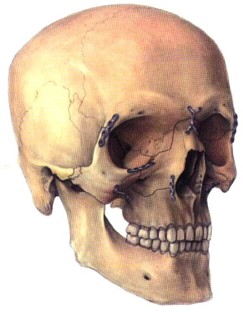
  • Direct fixation
  • Transosseous wiring at fracture sites
  • Frontozygomatic sutures
  • Infrorbital margin
  • Midline of the palate


Internal-wire suspension
  • Circumzygomatico-mandibular
  • Infraorbital border-mandibular
  • Frontomandibular
  • Pyriform fossa-mandibular


Support via the maxillary sinus by filling materials
  • Ribbon gauze
  • Balloon
  • Folly catheter
  • Polyethylene material
Length of the hospital stay will depend on a number of factors including:
  • Presence of other injuries
  • Age and medical status of the patient
  • Severity of the injury
  • Technique employed in the reduction and fixation of the fracture
  • Presence or absence of medical or surgical complications
  • Social circumstances of the patient